Massive Weight Loss Body Contouring: Extended Abdominoplasty Guide
8/5/2026
The patient: A 53-year-old Latina woman lost 120 pounds after diet, exercise, and RNY gastric bypass surgery (2019). She presented with severe excess skin of the abdomen and lateral torso (“side boob”). She also did not like the shape and position of her breasts. Her primary concerns were chronic rashes under her skin folds, difficulty exercising, and dissatisfaction with her appearance despite maintaining her weight loss.
Chief Complaint: “I have chronic rashes under my abdomen and pubic region. I have to use powder under my abdomen every day. I have difficulty buying bras and getting my “side boob” into my bra. I hate my body shape after losing 120 pounds.”
Weight History: The patient’s maximum weight was 340 pounds. The patient’s minimum weight was 230 pounds.
Previous Surgery: RNY gastric bypass surgery
Medical History: Morbid obesity (resolved)
Smoking: Remote history of vaping
Medications: None
Functional Symptoms: difficulty exercising and buying clothes
Physical Examination: 5'6", 250 pounds, BMI 40.4
Skin quality of abdomen: Poor with significant stretch marks above and below the umbilicus.
Skin quality of bilateral breasts: Poor with significant stretch marks. Skin quality of bilateral lateral torso: poor with significant stretch marks
Degree of laxity: Extreme abdominal skin laxity. Moderate breast skin laxity. Moderate lateral torso skin laxity.
Scars: Laparoscopic scar consistent with prior RNY gastric bypass. No hernias.
Muscle separation: 6 cm of rectus abdominis diastasis.
Lymphedema; none
Skin redundancy: extreme of abdomen, moderate of breasts and lateral torso
Breast: Bilateral grade 3 ptosis. Bilateral large areolar complexes. Sternal notch to nipple areolar complex measured 42 cm on right and 40 cm on left. Current bra 48 D
Diagnosis:
1) Massive weight-loss deformities of abdomen, breasts, and lateral torso.
2) Large abdominal pannus
3) Cutis pleonasmus of abdomen, bilateral breasts, and lateral torso.
4) Dermatochalasis of abdomen, bilateral breasts, and lateral torso.
5) Rectus abdominis diastasis
6) Bilateral grade 3 breast ptosis
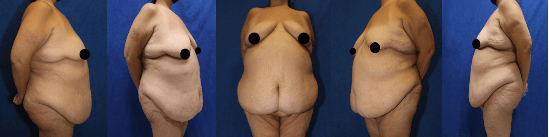
Figure 1: Pre-operative pictures of a 53 year old female patient who was 5'6" and 250 pounds. Her maximum weight was 340 pounds. She had lost 120 lbs. Pictures depict excess skin and fat of the abdomen, bilateral disproportionate breasts, bilateral breast ptosis, and excess skin and fat of the lateral side “boob.”
Why Surgery Was Recommended
Abdomen:Liposuction alone would remove fat, but would worsen skin redundancy. Because the patient had severe horizontal abdominal skin excess extending from above the umbilicus to below the supra-pubic region, and past bilateral superior iliac crests, an extended abdominoplasty was recommended. Because she rectus diastasis, rectus abdominis muscle repair was recommended.
Breasts: This patient had Grade 3 ptosis of bilateral breasts. She was content with her breast volume. Therefore a breast lift would be the best choice to achieve a lift without breast volume change. An anchor lift with an inferior based pedicle would yield the best result.
Lateral Torso: The patient had excess skin and fat of the lateral torso. Liposuction alone would remove fat,but would worsen skin redundancy. Therefore lateral torso skin needs to be excised. A oblique torsoplasty would probably yield the best result and can be extended from the anchor mastopexy incision.
Surgical Plan
Procedures Performed: extended abdominoplasty, bilateral anchor mastopexy, and bilateral lateral torsoplasty
Why Combined These Surgeries Were Combined: These surgeries were combined to minimize anesthesia time and recovery periods. The surgeries could have been staged. However, staging these surgeries into two or three parts would increase cumulative time under anesthesia, recovery time, time off work, and expense.
Expected Operating Time: 7 to 8 hours
Positioning: Supine
Incision Design And Preoperative Markings: An extended horizontal abdominoplasty was marked from the right superior iliac crest, under the mons pubis to a point 7 cm above the clitoris, to a point on the left superior iliac crest,. A proposed resection line was marked from the right superior iliac crest, above the umbilicus, and to the left superior iliac crest. An anchor mastopexy was marked in the infra-mammary fold with an 8cm inferior based pedicle. A new nipple areolar complex position was marked 20 cm from the sternal notch. 8cm vertical limbs were marked the remove the lateral most extent of the areola. The bottom of the 8 cm vertical limbs were joined horizontally with the infra-mammary fold incisions. An oblique torsoplasty was marked by extending the lateral portion of the anchor mastopexy in the infra-mammary fold. Tissue dissection will be carried cephalad and an elliptical area of skin and fat will be resected.
The Surgery: Extended Abdominoplasty
Pictures were taken and marking were made with a female chaperone. An IV was initiated. The patient was examined by a nurse and anesthesiologist. The patient was brought to the operating room, underwent IV sedation and general anesthesia The abdomen and bilateral breasts were then prepped with Betadine and draped in a routine sterile fashion. The proposed incision was then locally infiltrated with approximately 20 cc of 1% lidocaine with 1:200,000 of epinephrine. After waiting a full 7 minutes for effects, a #10 blade scalpel was used to make an incision in the abdomen, through the skin, through the fat, down to the anterior rectus fascia. Tissue dissection was carried cephalad to the umbilicus. The umbilicus was then cored out with the use of a #15 blade scalpel. A healthy 1-cm vascular stalk was left around the perimeter of the umbilicus. Tissue dissection was carried to the bilateral subcostal margins and the xiphoid centrally. Minimal bleeding was controlled with electrocautery. The wound bed was copiously irrigated with the use of normal saline and antibiotic solution and blotted dry. Minimal bleeding was controlled with electrocautery. The rectus abdominis musculature was then locally infiltrated on each side with the use of approximately 20 cc of 0.25% Marcaine with epinephrine. The rectus abdominis musculature was then medialized with the use of multiple interrupted figure-of-eight permanent sutures placed from the xiphoid to the umbilicus and then from the umbilicus to the suprapubic region. A second layer with a running permanent suture was also run from the xiphoid to the umbilicus and then from the umbilicus to the suprapubic region. Again, the wound bed was copiously irrigated and blotted dry. Minimal bleeding was controlled with electrocautery. Two #10 flat-fluted JP drains were placed at the base of the wound and brought out through the mons region and anchored with the use of a 3-0 permanent suture ties placed in a U-trapped door type stitch. On-Q pain pump was also inserted and placed on top of the rectus muscle and brought out inferiorly and attached to the JP drains with the use of Tegaderms. The proposed resection of the abdominal wall was confirmed 3x with the use of towel clamps before direct excision was accomplished. This was done with a #10 blade scalpel. Minimal bleeding was controlled with electrocautery. Absorbable interrupted sutures were placed in Scarpa’s fascia and the deep dermis. Next, a running absorbable suture was placed in the subcuticular layer. Any areas of skin discrepancy were then closed with the use absorbable sutures. The two #10 flat-fluted JP drains were brought out anteriorly and anchored with 2-0 permanent sutures placed in a U-trapdoor type fashion. A small stab incision was made at the intersection line from the xiphoid to the mons and another line perpendicular to this along the bilateral superior iliac crest. A 2-cm stab incision was made and an elliptical incision was created with the use of a #15 blade scalpel. The umbilicus was brought out and anchored out to 3, 6, 12, and 9 o’clock with the use of 3-0 absorbable sutures placed in an interrupted buried fashion. A perimeter circumferential absorbable suture was run around the perimeter of the umbilicus.
The Surgery: Bilateral Breast Reduction And Anchor Mastopexy
Attention was then focused on the bilateral breast mastopexy. The patient’s breasts were re-prepped with Betadine and re-draped in a routine sterile fashion. The patient was then elevated on the table and a temporary mastopexy and breast reduction pattern was completed with the use of staples. The aforementioned procedure was done on the contralateral side. The margins of the mastopexy were outlined and the staples were removed. The intervening skin was removed full thickness from epidermis to pre-pectoral fascia with the use of a #10 blade scalpel. The patient was then placed in a supine position. The mastopexy was then completed with the use of deep interrupted 2-0 absorbable sutures. This was then followed by running 3-0 absorbable suture. Laterally, the incision was carried from the infra-mammary fold posteriorly. the axillae. Closure was completed with interrupted, deep 2-0 PDS and 2-0 Monocryls, and then, a running 4-0 Monocryl. The nipple-areolar complex was brought out at the dome of the breast after a small round de-epithelized area was made at the pinnacle of the breast. This was then anchored at 12, 6, 3, and 9 o’clock with the use of 3-0 Monocryl absorbable sutures. A running circumferential periareolar permanent suture was placed around the perimeter of the nipple areolar complex. The procedure was done completely on the contralateral breast. At the completion of the procedure, the breasts were soft and no evidence of any ischemic compromise. The nipple-areolar complexes were both pink and intact.
The Surgery: Bilateral Oblique Torsoplasty
The proposed incision was locally infiltrated with approximately 40 cc of 1:100,000 of epinephrine. Next an incision was made from in the infra-mammary fold posteriorly. Tissue dissection was carried cephalad. The flap was then pulled caudally, and the proposed resection was confirmed 3 times with towel clips. Strict care was taken to avoid lateralization of the nipple areolar complexes. After confirmation, the ellipse of skin and fat was directly excised with a #10 blade. Minimal bleeding was controlled with electrocautery. The wound was irrigated with antibiotic solution and blotted dry. A #10 flat-fluted JP drains were placed on each side and attached to the skin with 2-0 permanent suture placed in a U-trap door type stitch. Scarpa’s fascia and deep subcutaneous tissue was then re-approximated with interrupted 1-0 and 0 absorbable sutures. A running 2-0 and 3-0 absorbable suture was placed in a subcuticular fashion to seal the wound. The procedure was performed on the contralateral side. The patient was wiped clean and dry of any blood and Betadine. Incisions were covered with 2 x 2 and secured with tape. A compression garment was placed. The patient was reversed from anesthesia, extubated, and taken to the recovery room in stable post-operative condition. The patient was monitored in the recovery center until their vital signs were stable.
The surgery took about 7 hours under general anesthesia.
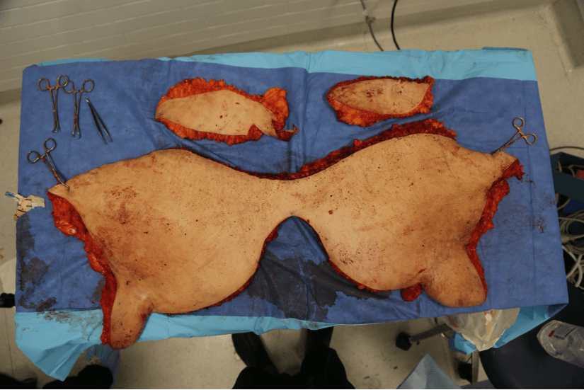
Figure 2: Picture of a 21lbs specimen of excess skin and fat taken form the abdomen.
The Specimen: 21 pounds was removed from the abdomen. Two pounds were removed from each lateral torso. Total weight removed was 25 pounds.
The Recovery: For the first two weeks, the patient was instructed to do minimal activity. She did 20 steps every day but otherwise was in a recovery bed. During her second two weeks, activity was increased to 40 steps twice a day. By the fourth week, she could do unlimited walking.
Pain: Postoperative pain was controlled with a On-Q lidocaine pain pump as well as oral medication specifically Percocet and Flexeril.
Medications: Patient was prescribed antibiotics. She continue the antibiotics while the drains were in place to minimize infection. Patient was given a stool softener to minimize constipation associated with narcotics. She was also prescribed a muscle relaxant (Flexeril) for pain control.
Position: For the first two weeks, she was encouraged to recover in a recliner. The recliner was angled at 45° of back flexion. Her arms were positioned out to the sides. When she walked, she was encouraged to bend at the waist and to use a walker.
Drains: Two drains were placed in the lower abdomen. These drains were removed when the drain output was less than 20 cc per day per drain for two consecutive days. The drains were removed around post-operative day #18.
Garments: Initially, the breasts were wrapped with a loose BIAS bandage. The abdomen was wrapped with abdominal binder. She was encouraged to wear the abdominal binder 24 hours a day for the first six weeks. She wore BIAS bandages on the breast for the first four weeks. Once the incisions were healed, she was then converted into a compression upper body garment.
Scars: At approximately six weeks, the scars were covered with Biocorneum silicone scar cream. On top of the Biocorneum, silicone tape was applied.. Gel Zone silicone strips are highly encouraged. These were applied every day for 12 hours. After 12 hours she was instructed remove the silicone cream and strips.
Nutrition: 100 grams of protein a day. This goal was achieved with Premier Protein shakes.
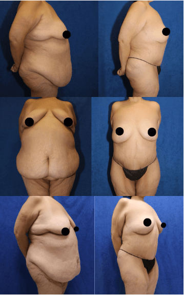
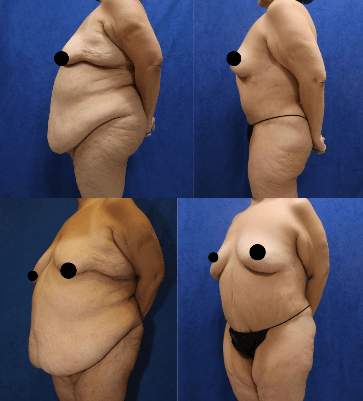
Figure 3: Post-operative pictures of the same patient above. She underwent extended abdominoplasty, bilateral breast reduction and anchor mastopexy, and bilateral oblique torsoplasty
The Results:
Abdomen: Much of the redundant excess abdominal skin and fat are gone. Due to the rectus tightening and removal of excess skin and fat, her abdomen is much flatter. After the surgery, her rashes resolved.
Breast: Her breasts are more proportional to the rest of her body. The rashes under her breasts are gone. Shoulder bra-strap pain and grooves gone. She can clothes and bras that fit.
Lateral torso: Her side boob is gone. Fittings for bras are much easier. Clothes fit better. She does not have to constantly adjust her bra to get the side of her breasts in to the bra.
The patient is very happy with her results. She can find clothing off the rack. She is able to exercise easier. Her rashes are gone. She is extremely satisfied that she underwent surgery with Dr. Katzen.